Thymosin Beta-4 Fragment (17-23)
Also known as: TB-500 Fragment · Ac-LKKTETQ · TB4 Frag
Thymosin beta-4 fragment (17-23) is a seven-amino-acid peptide, LKKTETQ, that corresponds to the actin-binding motif of thymosin beta-4. It is often supplied in an N-terminally acetylated form, Ac-LKKTETQ, which some researchers label TB-500. Laboratory studies have examined the motif in angiogenesis, wound healing and actin-binding assays. It is not an FDA-approved drug.
Last reviewed · Identity data matched to PubChem · 6 peer-reviewed sources
Key facts
- Class: synthetic fragment of thymosin beta-4; tissue-repair research peptide
- Length: 7 amino acids, LKKTETQ, matching residues 17 to 23 of thymosin beta-4
- Common form: N-terminally acetylated (Ac-LKKTETQ), sometimes labeled TB-500
- Formula C36H66N10O13; molecular weight 847.0 g/mol for the unacetylated peptide (PubChem CID 10169788)
- CAS 476014-70-7; PubChem synonym fequesetide
- Evidence base: in-vitro, animal and analytical studies; no clinical trials found
- Regulatory status: not FDA approved for any indication
Chemical Identity
| Molecular formula | C36H66N10O13 |
|---|---|
| Molecular weight | 847.0 g/mol |
| Monoisotopic mass | 846.4811 Da |
| CAS number | 476014-70-7 |
| PubChem CID | 10169788 |
| InChIKey | LVLWJWXFRFTMLK-JMWNXRNDSA-N |
| Compound type | Peptide |
| H-bond donors / acceptors | 14 / 16 |
| XLogP3 | -8.9 |
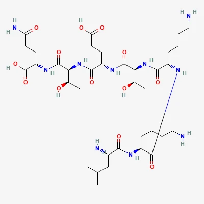
Structure notes: Commonly supplied with an N-terminal acetyl group (Ac-LKKTETQ). The PubChem record (CID 10169788) describes the unacetylated peptide with a free N-terminal amine and free C-terminal carboxylic acid.
What Is Thymosin Beta-4 Fragment (17-23)?
Thymosin beta-4 fragment (17-23) is the seven-residue peptide LKKTETQ, which reproduces the actin-binding motif of the 43-amino-acid protein thymosin beta-4 (Tβ4). Researchers have described this segment as the active site of Tβ4.[1]
Two forms appear in the literature. The unacetylated peptide, LKKTETQ, has been associated with actin binding, dermal wound healing, angiogenesis and skin repair.[1] The N-terminally acetylated form, Ac-LKKTETQ, is the molecule a 2024 analytical study calls TB-500, and that study noted that the biological effects of this acetylated form had not been documented before its work.[1]
Interest in the fragment follows from work on the parent protein. Thymosin beta-4 is the most abundant beta-thymosin in mammalian tissue and is regarded as the main G-actin-sequestering peptide; it is angiogenic and accelerates healing in dermal wound assays.[2] Studies of the 17-23 motif ask how much of that activity a short synthetic sequence can reproduce.
Chemical Structure and Molecular Identity
The fragment is a linear heptapeptide of standard L-amino acids: leucine, lysine, lysine, threonine, glutamic acid, threonine and glutamine. PubChem (CID 10169788) lists the unacetylated peptide with the molecular formula C36H66N10O13 and a molecular weight of 847.0 g/mol.
The acetylated form carries an acetyl group on the N-terminal leucine, which adds C2H2O, or about 42 Da, to the mass. Researchers working with Ac-LKKTETQ should therefore expect a higher mass than the PubChem value for the free peptide. Both forms end in a free C-terminal carboxylic acid on glutamine.
Two lysines and one glutamic acid give the peptide a net positive charge at neutral pH, and its computed XLogP of -8.9 reflects a highly polar, hydrophilic molecule.
Key identifiers
- PubChem CID: 10169788 (unacetylated LKKTETQ)
- CAS number: 476014-70-7
- InChIKey: LVLWJWXFRFTMLK-JMWNXRNDSA-N
- Synonyms: fequesetide, LKKTETQ, Tβ4(17-23), TB-500 fragment
Within full-length Tβ4, the 17-23 motif sits next to a basic lysine cluster at residues 14 to 19 (KSKLKK).[3]
Research History and Development
The 17-23 fragment emerged from efforts in the 1990s and 2000s to map which part of thymosin beta-4 binds actin and which part drives its biological effects. A 1996 mutational study using chemically synthesized full-length Tβ4 variants showed that the N-terminal region (residues 1-16) and a hexapeptide motif (residues 17-22) form separate structural entities that both contribute to actin binding.[4] Circular dichroism and activity data led the authors to a model in which the N-terminal part must adopt an alpha-helix, contacting actin through a hydrophobic patch at residues 6 to 12, while the motif contributes separate charged contacts.[4]
In 2003, researchers compared natural Tβ4, proteolytic fragments and synthetic peptides and reported that the seven-amino-acid actin-binding motif was essential for Tβ4’s angiogenic activity.[2] A 2006 proteomics study identified Tβ4 and thymosin beta-10 fragments in skin wound fluid and then tested LKKTETQ, the actin-binding domain the two proteins share, in wound models.[5] The fragments, with masses of 1565.7 and 1694.8 Da, were captured with capillary ultrafiltration probes and identified by mass spectrometry, the first identification of thymosin beta-10 in skin wound fluid.[5]
More recent work has turned to the acetylated peptide sold as TB-500. A 2024 study developed and validated an ultra-high-performance liquid chromatography and high-resolution mass spectrometry method to quantify Ac-LKKTETQ and its metabolites, using synthesized reference standards.[1]
Mechanisms Studied in Research
The fragment is studied mainly as an actin-binding and cell-adhesion motif, with additional evidence pointing to purinergic receptor signaling in wound repair. Mechanistic claims rest on in-vitro work and a small number of animal experiments.
Actin binding
Mutational analysis identified charged and hydrophobic residues in both the N-terminal helix and the 17-22 motif that contact actin. Electrostatic contacts involving lysine 18 in the motif, and lysine 14 in the N-terminal helix, appeared important for binding. The residues critical for actin contact are conserved across the beta-thymosin family, and a similar pattern occurs in the C-terminal headpiece of villin and dematin.[4]
Cell adhesion and angiogenic signaling
In the 2003 study, adhesion of cells to Tβ4 was blocked by the seven-amino-acid peptide, identifying the motif as the major cell-binding site on the molecule. Adding soluble actin inhibited both adhesion and vessel sprouting.[2]
Purinergic receptors and nuclear transport
The 2006 wound study suggested that LKKTETQ’s effect on healing may be mediated by purinergic receptors.[5] Separately, experiments with Tβ4 variants carrying substitutions in the 17-23 motif found that nuclear localization of Tβ4 did not depend on its actin-binding ability or on the basic lysine cluster.[3] The authors proposed passive but regulated diffusion as the route by which Tβ4 shuttles into the nucleus.[3]
Key Findings From Published Studies
Published findings on the 17-23 fragment come from cell culture, chick embryo tissue, rodent wound models and rat metabolism studies; no human trials were found. Results are summarized by model below.
In-vitro and ex-vivo studies
- Endothelial migration and sprouting: in human umbilical vein endothelial cell migration assays and chick aortic arch sprouting assays, Tβ4 and its actin-binding motif showed near-identical activity, while peptides lacking any portion of the motif were inactive.[2]
- Skin wound healing: LKKTETQ significantly enhanced skin wound healing in vitro and in vivo.[5]
- Fibroblasts: neither Ac-LKKTETQ nor its metabolites were cytotoxic, and only the metabolite Ac-LKKTE showed significant wound-healing activity compared with control.[1]
- Ovarian cancer cells: full-length Tβ4 and three Tβ4-derived peptides, including Tβ4(12-26), which spans the motif, and peptides without an actin-binding site, all increased migration, invasion and proliferation of SKOV3 cells and up-regulated RACK-1 protein.[6]
Metabolism in rats and human serum
The 2024 study examined Ac-LKKTETQ metabolism in human serum, several in-vitro enzyme systems and urine from treated rats. Ac-LK was the primary metabolite, reaching the highest concentration in rats during the first 6 hours, while Ac-LKK was a long-lived metabolite detected up to 72 hours. The authors concluded that wound-healing activity previously reported for TB-500 may be due to the metabolite Ac-LKKTE rather than the parent peptide.[1]
Because acetylated and unacetylated forms differ in mass and reported activity, researchers typically confirm the form with HPLC and mass spectrometry certificates of analysis.
Because acetylated and unacetylated forms differ in mass and reported activity, researchers typically confirm the form with HPLC and mass spectrometry certificates of analysis.
Regulatory and Research Status
Thymosin beta-4 fragment (17-23) is not approved by the FDA for any indication, and no clinical trials of it were found in PubMed as of October 2026. Its literature consists of structural, in-vitro, animal and analytical studies.
Several open questions remain. The acetylated form has been far less studied than unacetylated LKKTETQ, and the 2024 metabolism data suggest that a metabolite may account for at least part of its reported wound-healing activity.[1] The ovarian cancer cell findings also show that Tβ4-derived peptides can stimulate migration and invasion of tumor cells in vitro.[6]
The fragment is supplied as a research chemical for laboratory use. Because the name TB-500 is applied both to this heptapeptide and to full-length thymosin beta-4, sequence and mass data are the most reliable way to establish what a given sample contains.
Frequently Asked Questions
Is TB-500 the same as thymosin beta-4 fragment 17-23?
Sometimes. A 2024 analytical study uses TB-500 to mean Ac-LKKTETQ, the acetylated 17-23 fragment of thymosin beta-4. The same name is also widely applied to full-length thymosin beta-4, a 43-residue peptide with a molecular weight near 4963 g/mol. Mass spectrometry easily distinguishes the two, since the fragment is under 900 Da.
What is the molecular weight of LKKTETQ?
PubChem lists unacetylated LKKTETQ (CID 10169788) with a molecular weight of 847.0 g/mol and the formula C36H66N10O13. The N-terminally acetylated form, Ac-LKKTETQ, adds an acetyl group of C2H2O, so its mass is about 42 Da higher. Researchers should check which form a certificate of analysis reports.
Why is LKKTETQ called the actin-binding motif?
Mutational studies of thymosin beta-4 identified residues 17 to 22 as a separate structural unit that contacts actin, with lysine 18 forming an important electrostatic contact. A 2003 study showed the seven-residue LKKTETQ sequence was essential for thymosin beta-4's angiogenic activity and was the main site through which cells adhered to the protein.
Is thymosin beta-4 fragment FDA approved?
No. Thymosin beta-4 fragment (17-23) is not approved by the FDA for any use, and no clinical trials of it were found in PubMed as of October 2026. Its published literature is limited to structural, cell culture, animal and analytical chemistry studies. It is supplied as a research chemical for laboratory use.
What does the research on the TB-500 fragment show?
In laboratory studies, LKKTETQ matched full-length thymosin beta-4 in endothelial migration and vessel sprouting assays and enhanced skin wound healing in vitro and in vivo. A 2024 rat and serum study found that the acetylated form is metabolized to shorter peptides such as Ac-LK and Ac-LKK, and that the metabolite Ac-LKKTE, not the parent peptide, showed wound-healing activity in fibroblasts.
How is the 17-23 fragment different from full-length thymosin beta-4?
The fragment contains only seven of thymosin beta-4's 43 amino acids. It retains the actin-binding motif but lacks the N-terminal helix that also contributes to actin binding. Its molecular weight is roughly one sixth of the full peptide's, and it has been studied far less, with no clinical trials found.
References
- Rahaman KA, Muresan AR, Min H, et al. Simultaneous quantification of TB-500 and its metabolites in in-vitro experiments and rats by UHPLC-Q-Exactive orbitrap MS/MS and their screening by wound healing activities in-vitro. Journal of chromatography. B, Analytical technologies in the biomedical and life sciences 2024. PMID 38382158 · DOI
- Philp D, Huff T, Gho YS, et al. The actin binding site on thymosin beta4 promotes angiogenesis. FASEB journal : official publication of the Federation of American Societies for Experimental Biology 2003. PMID 14500546 · DOI
- Zoubek RE, Hannappel E. Subcellular distribution of thymosin beta4. Annals of the New York Academy of Sciences 2007. PMID 17567947 · DOI
- Van Troys M, Dewitte D, Goethals M, et al. The actin binding site of thymosin beta 4 mapped by mutational analysis. The EMBO journal 1996. PMID 8617195
- Huang CM, Wang CC, Barnes S, et al. In vivo detection of secreted proteins from wounded skin using capillary ultrafiltration probes and mass spectrometric proteomics. Proteomics 2006. PMID 17001601 · DOI
- Yoon HJ, Oh YL, Ko EJ, et al. Effects of thymosin β4-derived peptides on migration and invasion of ovarian cancer cells. Genes & genomics 2021. PMID 34170491 · DOI
Related Compounds
This profile summarizes chemical data and peer-reviewed literature for educational and laboratory-research reference. It is not medical advice, and nothing here describes or recommends use in humans or animals. How we source and verify data.